
There are two common questions we get during a hands-on workshop regarding fetal breathing during a biophysical profile exam.
- How do you identify fetal breathing?
- How long do you have to see it?
Let’s tackle the first question. In order to evaluate fetal breathing, you will need to image the chest and diaphragm. I like to do this in a sagittal plane. Once you have established fetal lie, position your transducer in a sagittal plane on the fetus. You will need to sweep (remember the paint brush motion) in order to move the spine or ribs out of the way. Once you have a nice clear image of the chest and diaphragm, sit there and wait. Depending on what side of the baby you are on, you may see liver, heart, and stomach as well.
See image below.
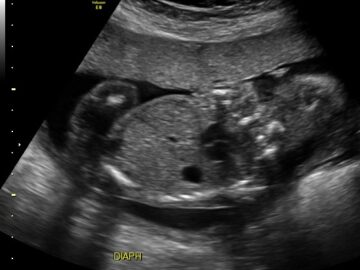
In this image, you can see the liver, stomach, part of the heart and the diaphragm. It is really nice if you can get the fetal spine down so you do not have to worry about shadowing. This is done by sweeping left to right on baby in a sagittal plane.
Here is another example.
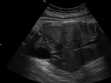
Here the baby is lying on it’s side. We are getting an anterior shot of the diaphragm. You can see the ribs, diaphragm, liver and fetal bladder. Once you achieve this image, rest your hand on mom’s belly gently to steady the probe. Like this:

Now wait. If you are doing this as part of a biophysical profile, check your time. I always start my BPP exams with fetal breathing. Most of the time the baby will move while I’m waiting and I can check that off. Also, don’t confuse maternal breathing with fetal breathing. Maternal breathing is slow and consistent. Maternal breathing moves the whole uterus. Fetal breathing is quick and specific to the diaphragm and liver moving.
See video below.
Remember that you need to see 30 seconds of consistent fetal breathing within a 30 minute window to give points for the biophysical profile.
For a complete review of the biophysical profile visit our online course page.