
RPOC Discussion
Retained products of conception or RPOC refers to the tissue that remains in the uterus after a pregnancy has ended. This tissue may be seen after abortion, miscarriage, vaginal delivery, or c-section. RPOC describes a broad category of tissues – it is usually placental but can be fetal as well. Retained products of conception are uncommon, but can cause a variety of complications including anemia, chronic pelvic pain, infection, sepsis, and scarring of the uterus or Asherman’s syndrome.
Symptoms of RPOC include heavy or irregular bleeding, passing clots, vaginal discharge, enlarged or tender uterus, fever, pain, infection, or a missed period. Patients are a greater risk of having RPOC if the following apply to them: placenta accreta, advanced maternal age, assisted delivery (vacuum or forceps), second trimester delivery, failure to progress, nulliparity, uterine surgeries, or an irregular shaped uterus.
Typically testing will include a transvaginal ultrasound with color Doppler and an HCG. If the tissue is placental, the HCG will still be positive and may be high. Treatment includes the use of misoprostol, hysteroscopy, and/or D&C.
Case Study
Patient History
Patient presented as a 28-year-old G4P2 with a positive pregnancy test. LMP is noted to be July 17th. The patient had an elective abortion with the first dose of medication give in the clinic on September 7th and then had the subsequent doses at home on September 8th. The patient reported heavy bleeding and cramps for about 4 days and then just light spotting until September 16th. A transvaginal ultrasound was ordered and performed on October 8th. CPT code 76830 was used.
Transvaginal Ultrasound Images
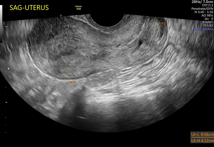
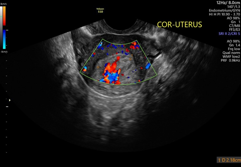
The sagittal uterus demonstrates a thickened and inhomogeneous endometrium.
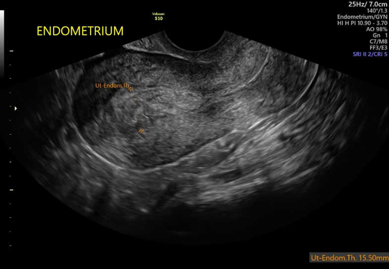
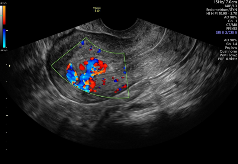
The lining of the uterus measured 15.5mm and is hyper-vascular.
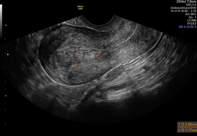
A well-defined collection of tissue is seen within the cavity that measures 2.9 x 2.2 x 1.2
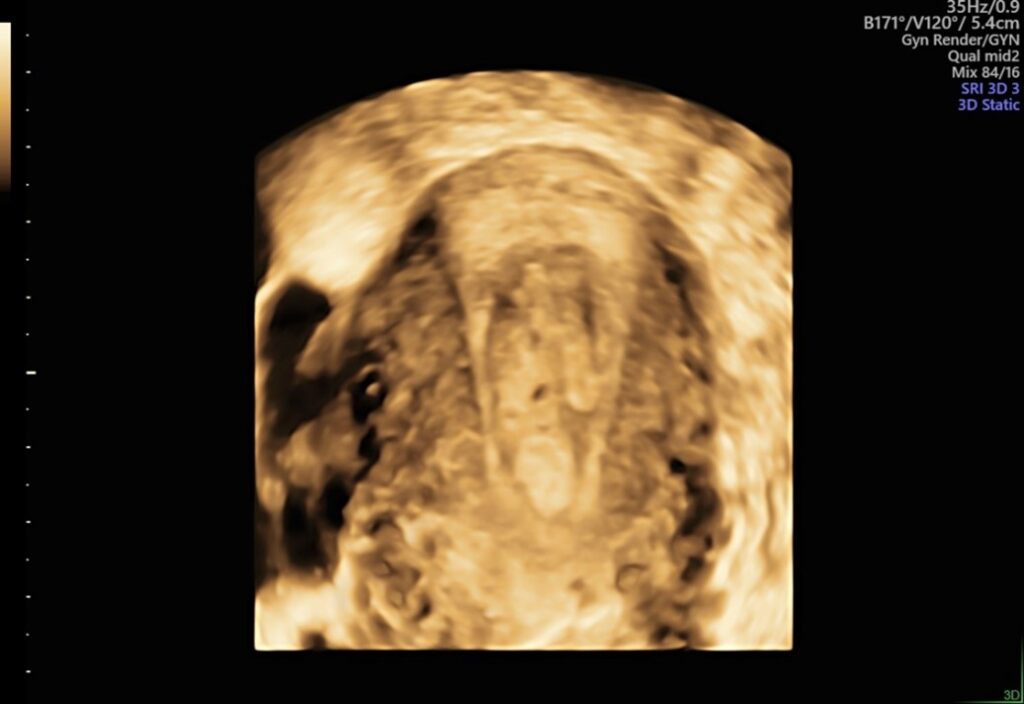
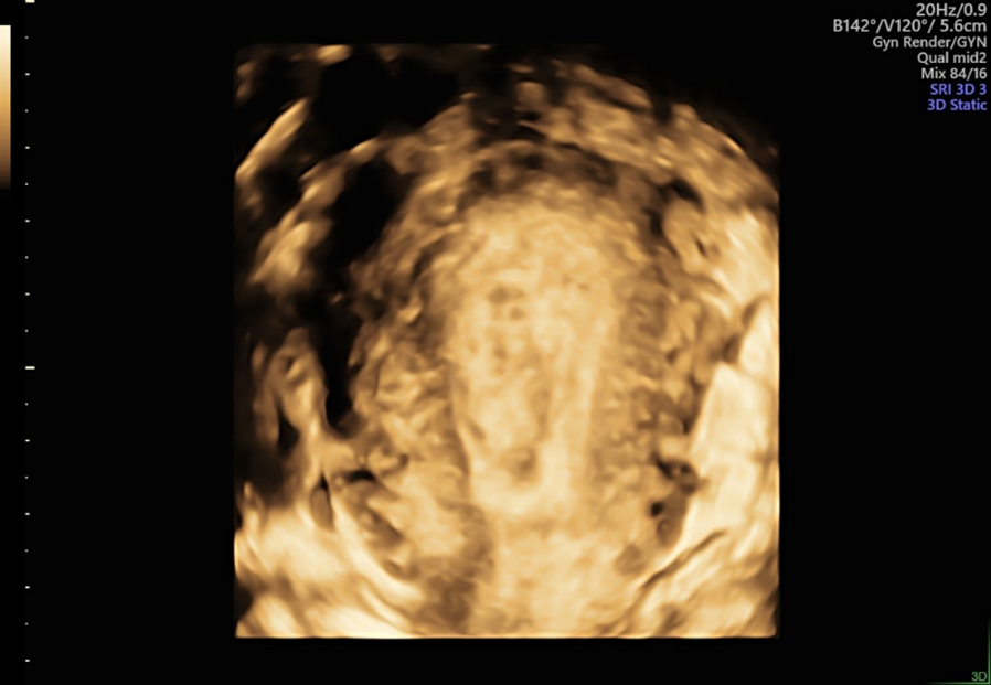
A 3D image of the cavity is obtained which also shows the collection of tissue.
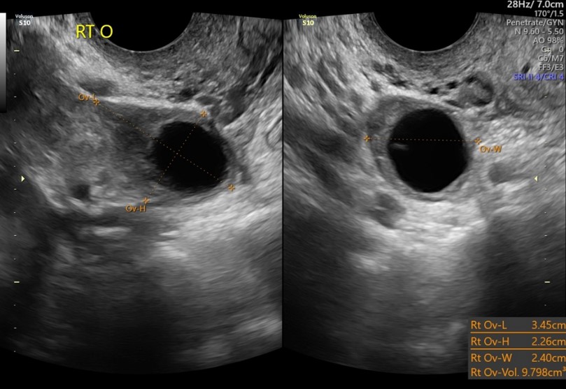
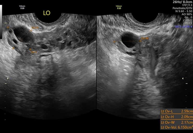
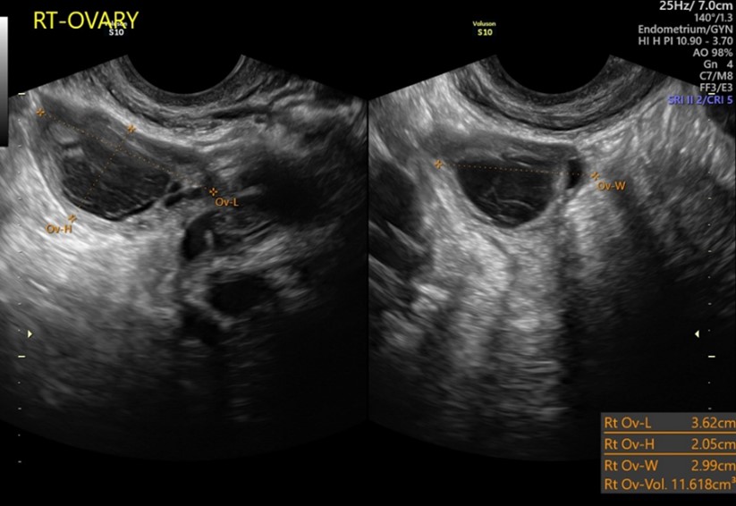
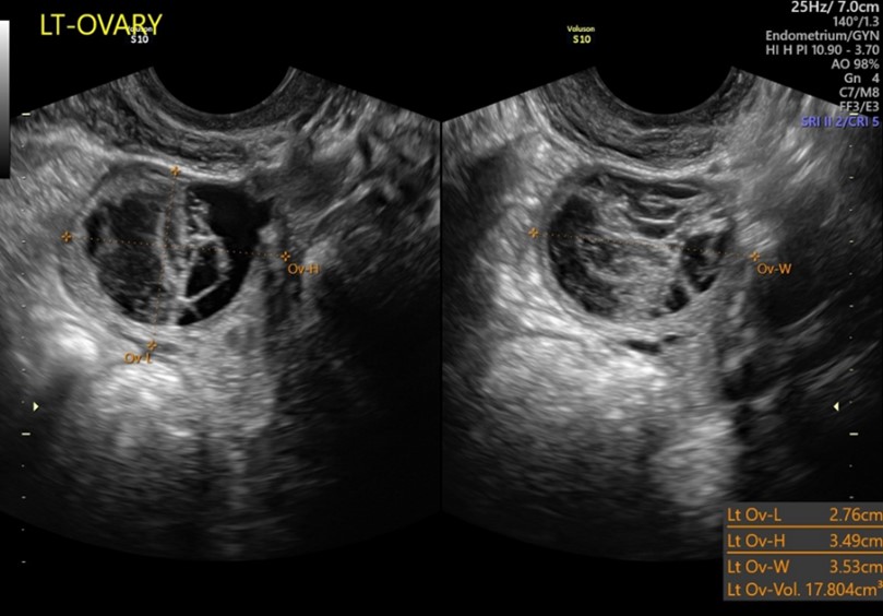
Both ovaries are evaluated on all GYN scans, and the ovaries appear normal.
Findings
The findings of the ultrasound were consistent with RPOC. The patient was prescribed misoprostol and scheduled to return on October 16th for follow up ultrasound. When the patient returned, she reported no additional bleeding or clots with the medication.
Follow Up Ultrasound Images
Notice that the follow up ultrasound appears very similar to the prior scan since the patient reported no bleeding after taking the misoprostol.
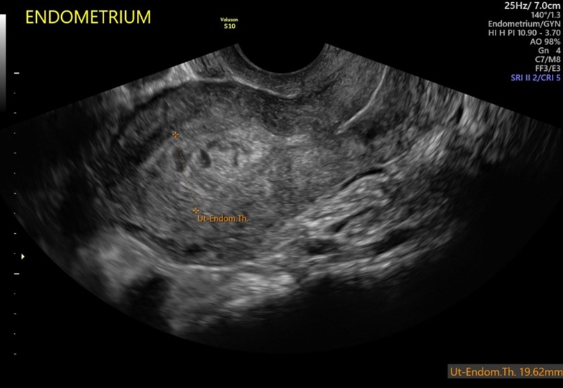
The sagittal uterus still demonstrates a thickened and inhomogeneous endometrium.
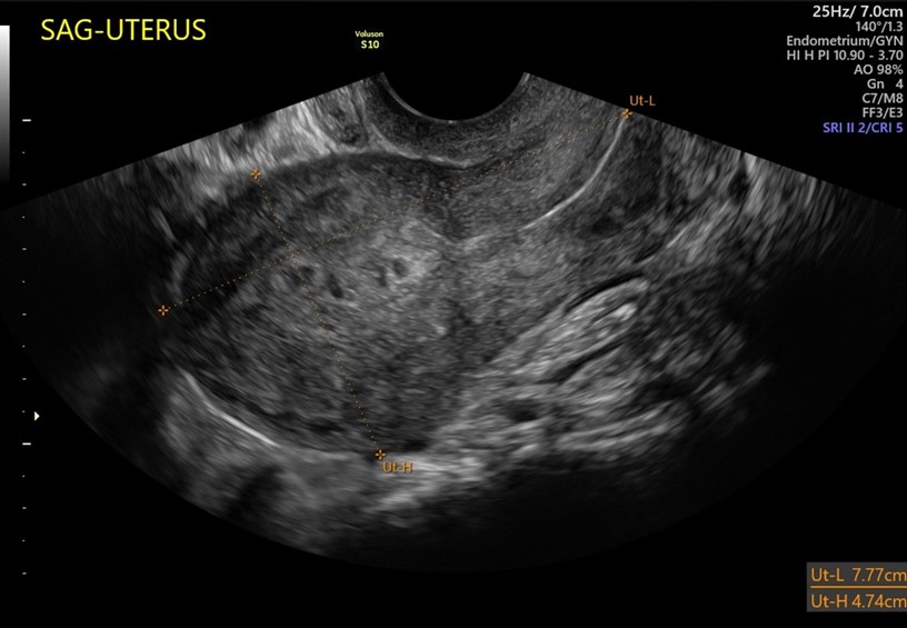
The endometrium measured 19.62mm, which is slightly thicker than previously documented and the hypervascularity remains.
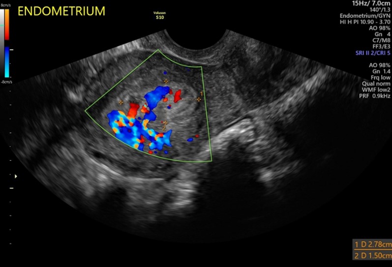
A well-defined collection of tissue is again seen within the cavity measuring 2.8 x 1.9 x 1.5cm – essentially unchanged from prior study.
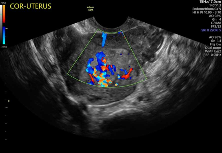
Again, note the intense hypervascularity.
Another 3D image of the cavity appears like the first, with the well-defined collection of tissue still visible in the cavity.
The ovaries were re-evaluated, and bilateral, small complex and hemorrhagic cysts were noted.
The ultrasound findings were unchanged. RPOC again noted in the cavity. The patient was given a second prescription of misoprostol, but two days later called back to report that she still was not bleeding. D&C has been scheduled.